Hoboken’s valves
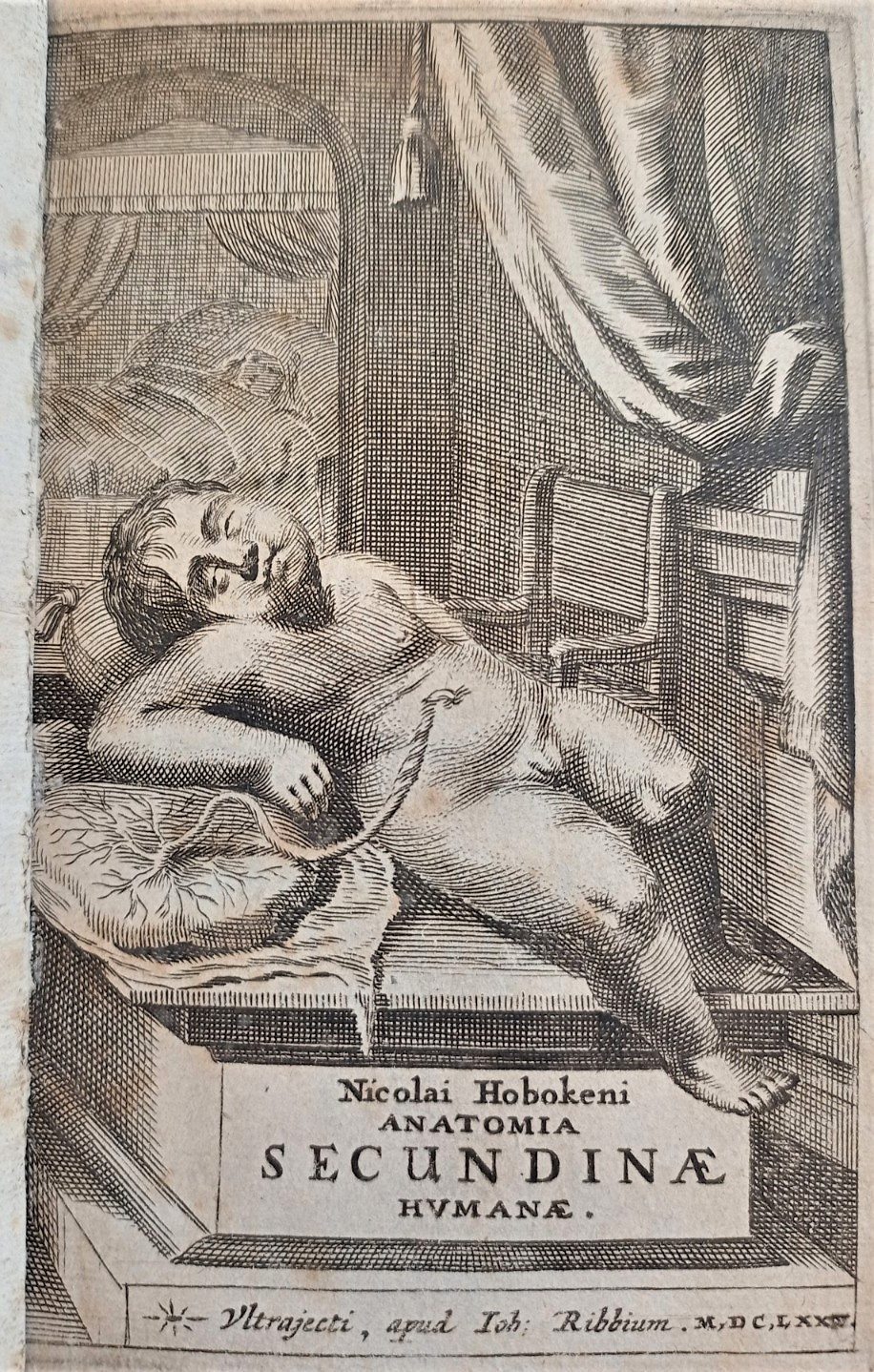
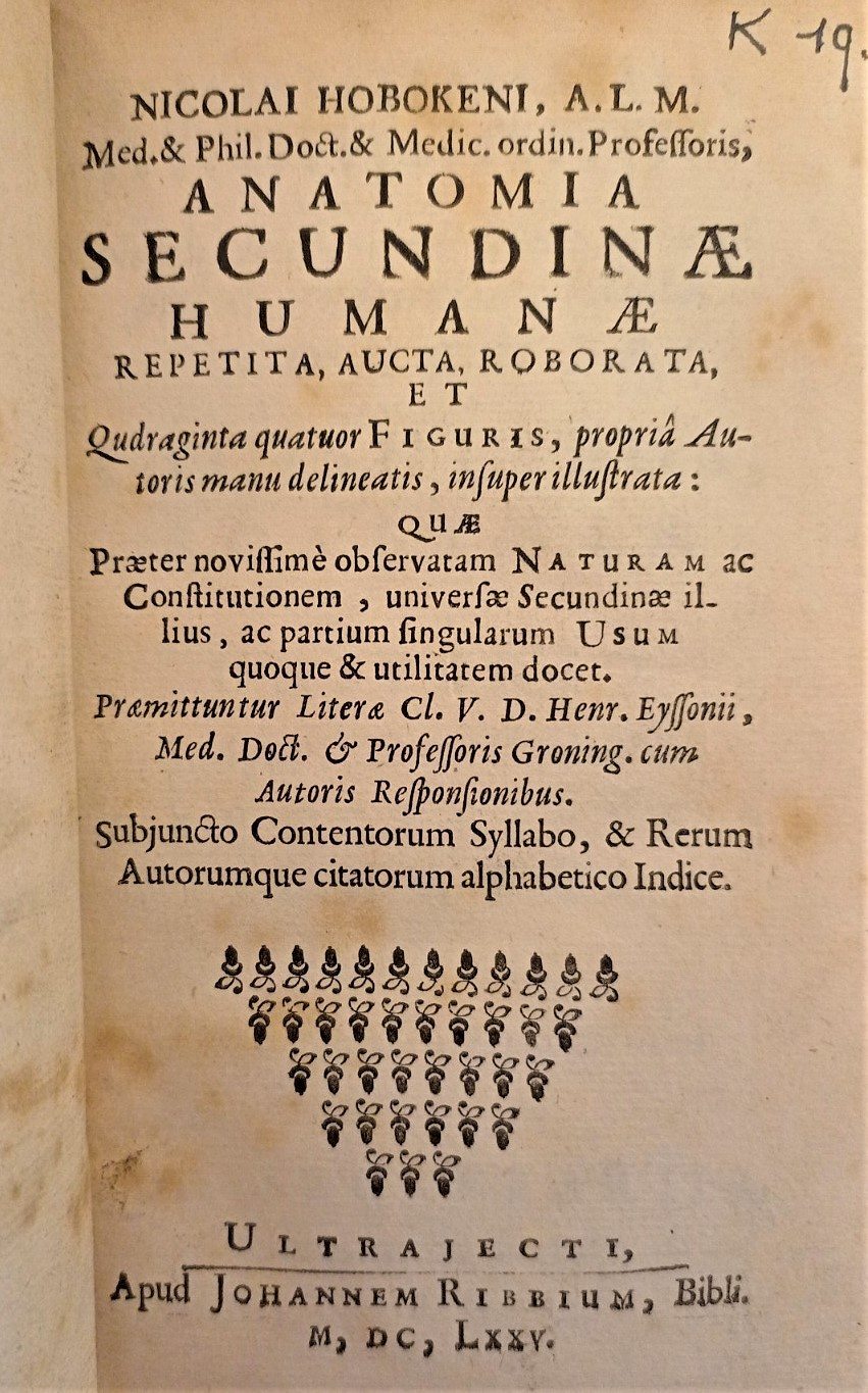
Anatomia secundinæ humanæ (Anatomy of the human afterbirth), by Nicolaas Hoboken. Printed in Utrecht by Johannes Ribbius, 1675. Lower Library, K.19.7
Nicolaas Hoboken (1632–1678) was a professor of medicine and mathematics, now best known in relation to his discoveries and theories about the umbilical cord. After the discoveries of Fabricius and Harvey, it had become clear that previous, Galenic beliefs about the flow of blood through the umbilical arteries could not be accurate.1
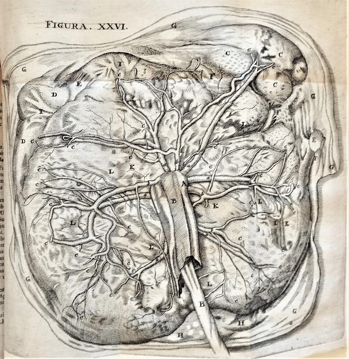
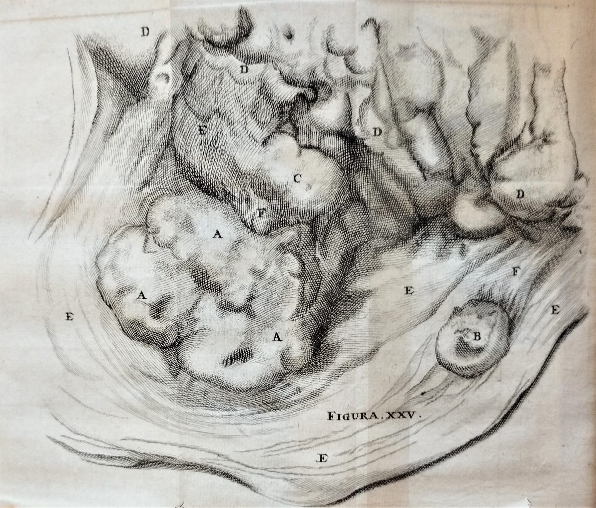
Hoboken was the first to illustrate the vessels of the umbilical cord, dissecting them carefully and then drawing the images for his text himself. He discovered folds inside the umbilical arteries, previously unknown, and theorised that they were valves to help with the movement of blood. These came to be known as the valves or nodes of Hoboken.2
Hoboken’s ‘valves’ have been the source of some controversy. It is now generally agreed that there are no valves in the umbilical arteries, but the exact nature and purpose of the folds which Hoboken identified have historically been the subject of some debate.3 It is now theorised that these are rings which contract in order to close off the arteries after birth.4
His study also covered the placenta, and fetal membranes, and was incredible in its detail, although he did mistakenly claim that there was no exchange of blood between the mother and the fetus.
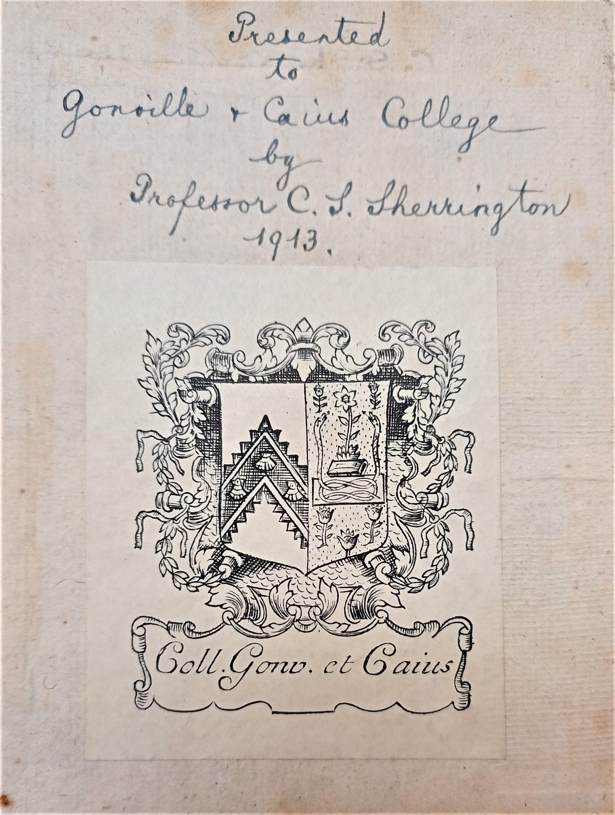
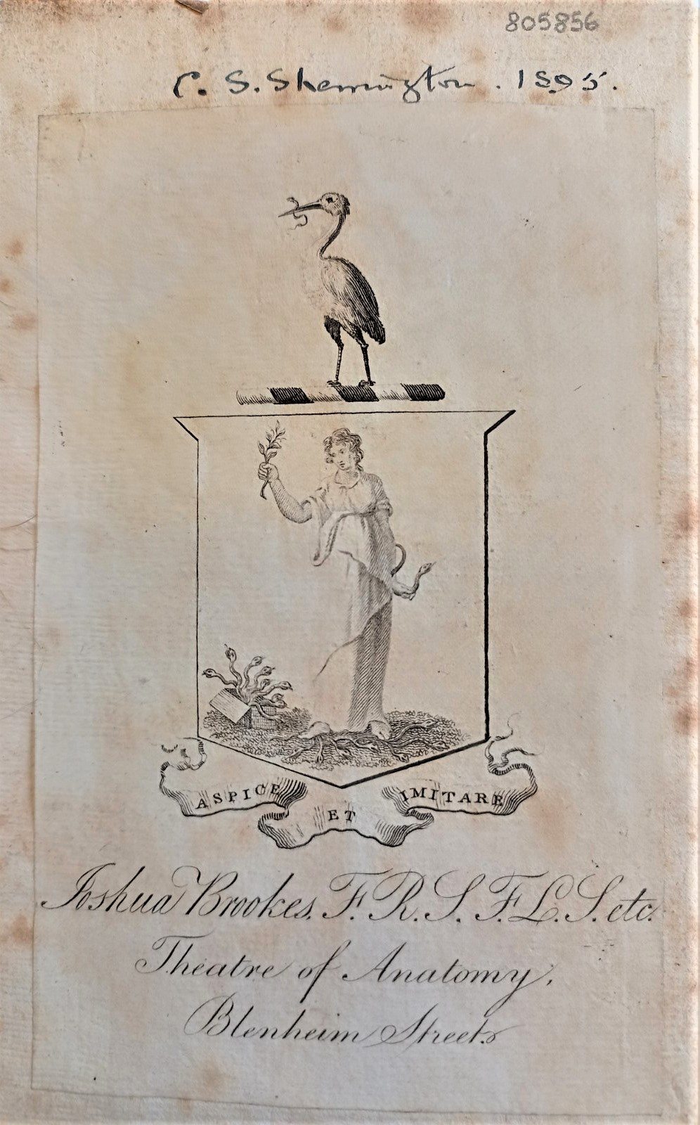
Our copy of this work was donated to Caius by C. S. Sherrington, an eminent neurophysiologist and a Caian. Sherrington went on to win the Nobel Prize in Physiology or Medicine in 1932.
Witness to the Black Death << Hoboken’s valves >> A working book for an idler
- Lawrence D. Longo and Lawrence P. Reynolds, Wombs with a View: Illustrations of the Gravid Uterus from the Renaissance through the Nineteenth Century (Cham: Springer International Publishing AG, 2016), 72.
- Thomas F. Baskett, ‘Hoboken, Nicolaas (1632–1678): Folds of Hoboken’, in Eponyms and Names in Obstetrics and Gynaecology, 3rd ed. (Cambridge: Cambridge University Press, 2019), 183–84.
- Longo and Reynolds, Wombs with a View, 72.
- G. Röckelein and A. Scharl, ‘Scanning Electron Microscopic Investigations of the Human Umbilical Artery Intima’, in Vichows Archiv 413, no. 6 (1988): 555–561.
